Contact-Free Biosignal Acquisition via Capacitive and Ultrasonic Sensors
R. Kusche, Fabian John, Marco Cimdins, and H. Hellbrück, IEEE Access, 8, 2020; DOI: 10.1109/ACCESS.2020.2995861; [PDF]
Abstract: Contact-free detection of human vital signs like heart rate and respiration rate will improve the patients’ comfort and enables long-term monitoring of newborns or bedridden patients. For that, reliable and safe measurement techniques are indispensable. The aim of this work is the development and comparison of novel ultrasonic and capacitive measurement setups, sharing a common hardware platform. Both measurement techniques that are implemented and compared are based on the detection of minor chestwall vibrations in millimeter ranges, due to geometrical thorax changes during respiration and heartbeat activities. After examining the physical measurement conditions and simulating the capacitive sensor, a problem-specific measurement setup is proposed. The system is characterized to be capable of detecting distance changes below 2 µm via the ultrasonic sensor and below 800 µm via the capacitive sensor. First subject measurements show that the detection of heart activities is possible under ideal conditions and exclusively with the proposed ultrasonic approach. However, the capacitive sensor works reliably for respiration monitoring, even when the subject is fully-clothed and covered with a blanket. The chosen ultrasonic approach is sensitive regarding minor changes of the reflecting surface and therefore has high uncertainty. In contrast, capacitive respiration detection is very reliable. It is conceivable that improvements in the capacitive sensor circuitry will also enable the detection of heart activities. The proposed ultrasonic approach presents current problems of this technique. In contrast to that, the unusual approach of capacitive sensing demonstrates a high potential regarding vital signs acquisition.
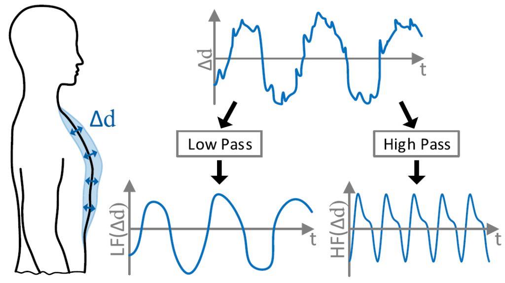
Figure 1: Principle of biosignal acquisition by analyzing geometrical changes of the chest wall during respiration and heart activities. The low-frequency components are considered to be caused by respiration and the high-frequency components are related to the heart activities.
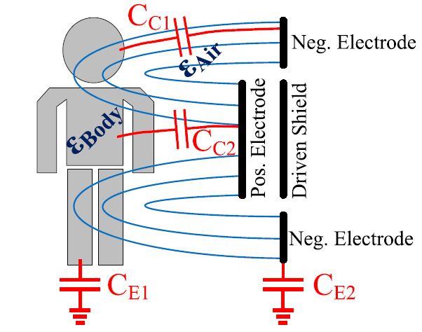
Figure 2: Principle of capacitive distance measurement. The circular sensor consists of an inner positive electrode and an outer negative electrode ring. An optional driven shield can be used for unidirectional field propagation. This principle is based on the different permittivity values of air and the human body. In addition, the parasitic capacitive coupling of the human body (CE1) and the sensor (CE2) affects this approach.
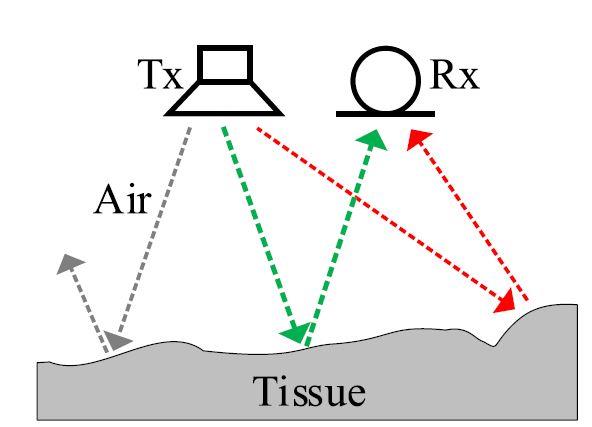
Figure 3: Simplified principle of ultrasonic distance measurement. The uneven surface of the reflecting material, in this approach human tissue, causes scattering effects. The green dashed arrow represents the desired path to determine the distance between the tissue and the transmitter (Tx)-receiver (Rx) setup.
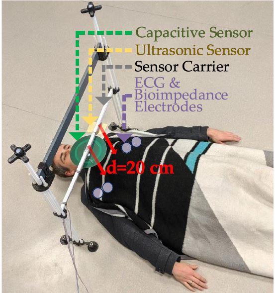
Figure 4: Measurement setup to acquire respiration and heart
activities simultaneously via both proposed approaches. For reference
measurements, additionally, the ECG and bioimpedance are measured
across the thorax. The air distance between the sensors and the subject is
20 cm. The first measurements are performed bare-chested. Afterward,
the procedure is repeated fully-clothed with a T-shirt, pullover, winter
jacket, and a blanket, as shown in this photograph.
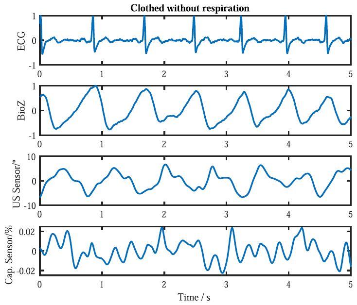
Figure 5: Measurement results from the bare-chested subject during
holding the breath for 5 seconds. In the ECG and the BioZ plots,
the expectable heart activity can be seen. The signal from the ultrasonic
sensor contains the heartbeat correlated information, as well. However,
it cannot be detected in the capacitive sensor signal.
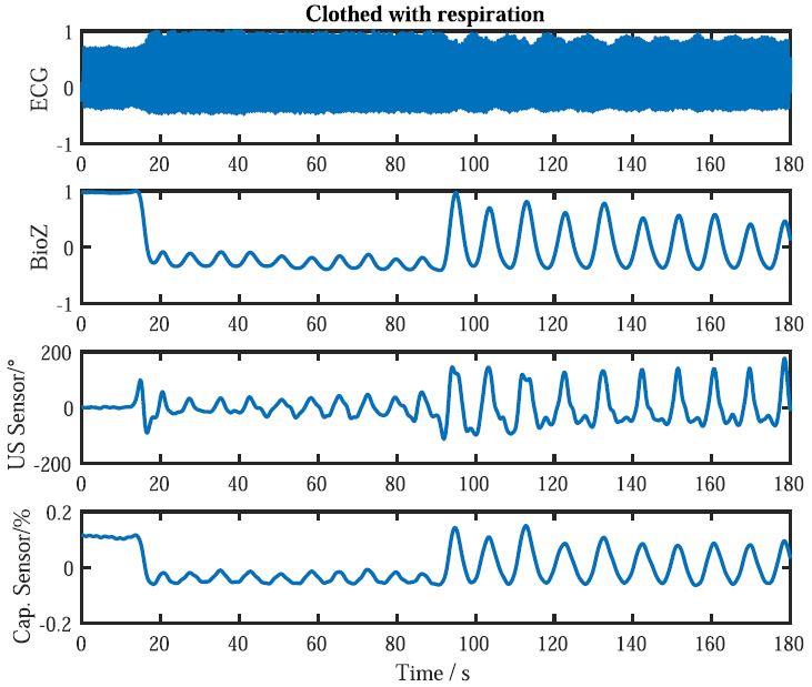
Figure 6: Measurement results from the fully-clothed subject during
holding the breath for 5 seconds.